
ADVANCED IMAGING
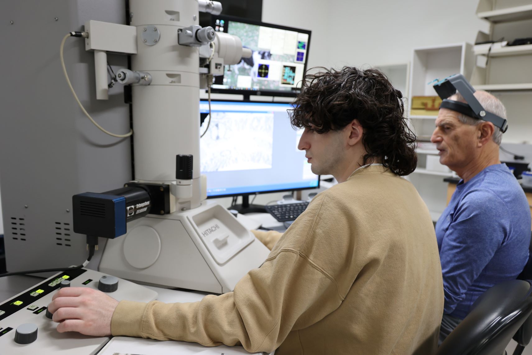
State-of-the-art scanning and transmission electron microscopes are used by students for teaching and research with faculty in the UW-Stevens Point Department of Biology. The Electron Microscopy Laboratory on the Haeni Chemistry Biology Building third floor features two different types of microscope.
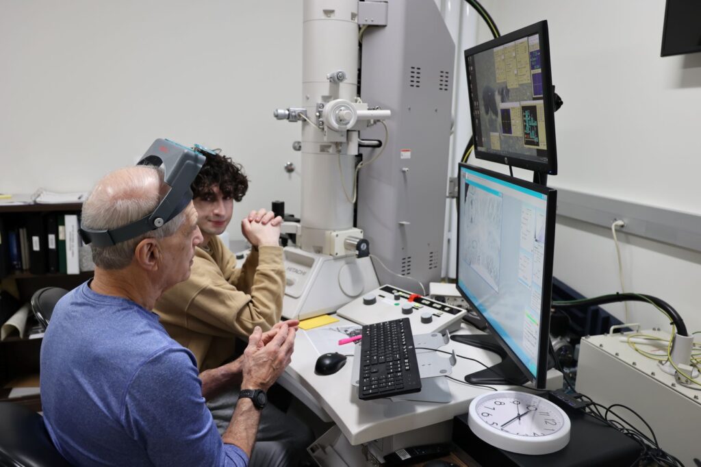
The powerful microscopes allow students to study at the molecular level. Undergraduates engaged in research projects at UW-Stevens Point work under the direction of faculty or in independent study.
Our facility is supervised by Professor of Biology Qiang Sun, Electron Microscopy Laboratory director.
Electron Microscope Facility
Transmission Electron Microscope
An Hitachi H-7500 transmission electron microscope (TEM) is used for looking at either sections of tissue or specially-prepared viruses or macromolecules mounted on 3mm grids. It has a magnification range of 700x to 600,000x, about 400 times better than the best light microscope.
Scanning Electron Microscope
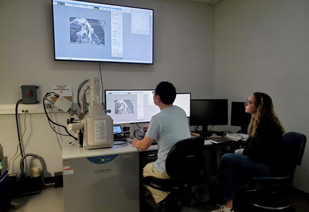
The second type of EM is an Hitachi S-3400 scanning electron microscope (SEM), for looking at whole, unsectioned samples such as insects or plants, up to the size of a softball. It has a useable magnification range of 10x to 50,000x. The SEM is also equipped with an X-ray detector that can determine which elements are found in alloy samples put into the microscope.
Research is a highly creative and challenging endeavor which allows students to apply the knowledge and skills learned in the classroom towards solving problems and developing new knowledge.
View Other Specialized EquipmentStudents are guided by a faculty mentor but work independently in the laboratory. They often present their work at the on-campus Letters and Sciences Undergraduate Research Symposium, at off-campus scientific conferences, and as co-authors on refereed journal articles. For some students, their research experience at UW-Stevens Point is a life changing experience as they decide to pursue graduate studies in chemistry or biochemistry.